


Looking towards the upcoming National Citizens Inquiry (NCI) hearing scheduled for August 18–20, 2026, where I am set to deliver expert forensic testimony on the emerging biosecurity and public health implications of Dr. John Fagan’s laboratory findings when testing synthetic milk, I am compelled to reflect on my previous appearances before this independent body. We must evaluate past data before we can project future risk.

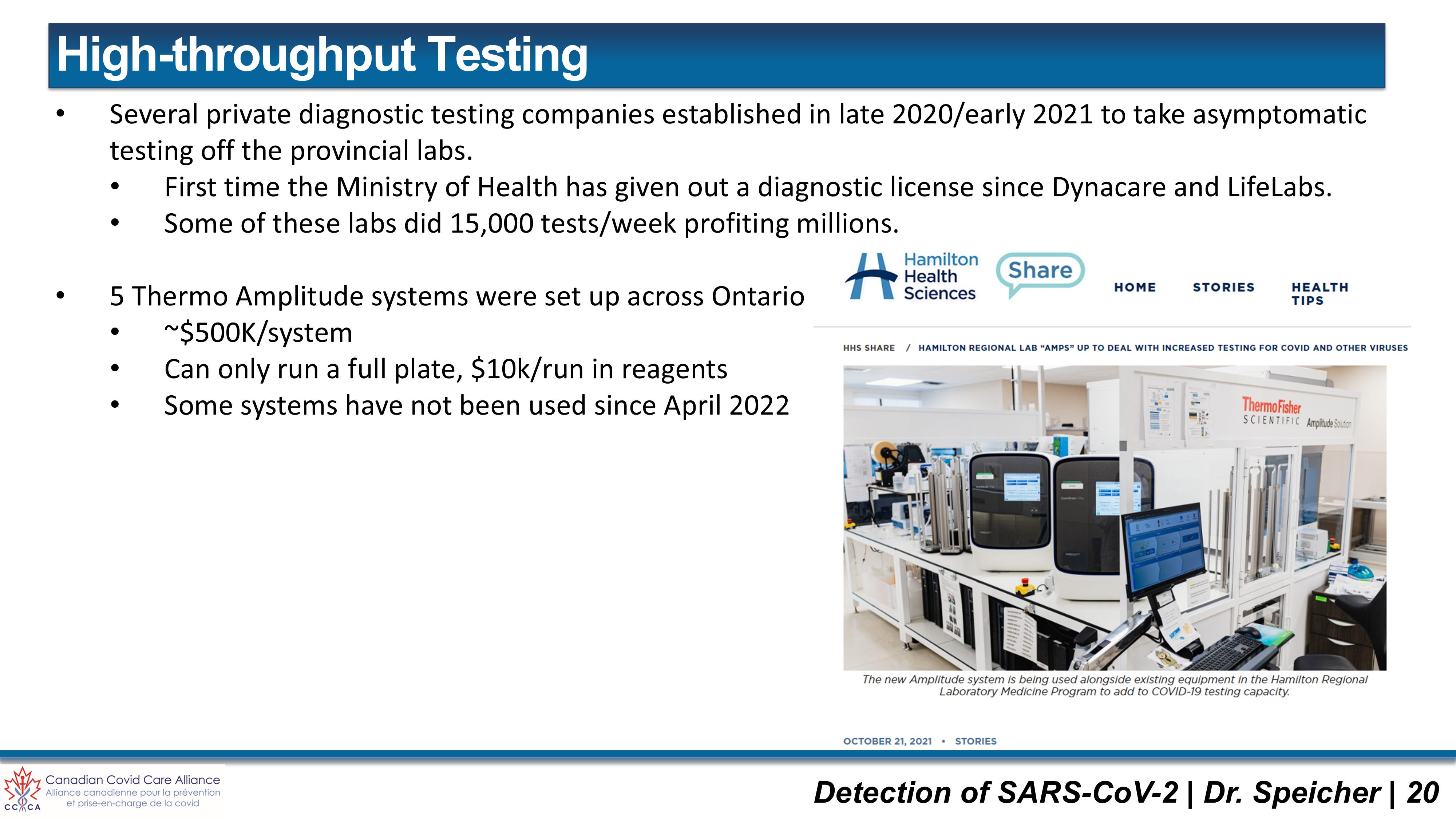
On May 18, 2023, I first took the stand at the NCI in Ottawa to address the foundational instrument of the entire pandemic response: the widespread clinical misuse of Polymerase Chain Reaction (PCR) testing. Having served as Laboratory Director for two high-throughput genomic testing facilities overseeing up to 15,000 PCR assays per week testing asymptomatic people, I detailed how an extraordinarily elegant laboratory technique was stripped of its clinical diagnostic context and weaponized to drive administrative fear. Subsequently, on March 7, 2025, I returned to the NCI to present our peer-reviewed laboratory findings detailing residual plasmid DNA and SV40 promoter-enhancer sequences contaminating modRNA vaccine platforms. It was this exact molecular tool—quantitative PCR (qPCR)—that enabled these genetic impurities to be detected and quantified. This definitive empirical evidence was then entered into the national record.
The Lineage of Truth: From a Spark to a National Record
When official channels deflect empirical data with bureaucratic procedure, independent scientific and legal professionals must assume the auditing responsibility. This mandate catalyzed the creation of the National Citizens Inquiry (NCI)—an independent initiative designed to establish an immutable legal and historical record of the pandemic era. Moving coast-to-coast, the NCI methodically organized its inquiry across three core pillars: Medical and Scientific Sovereignty, Legal and Constitutional Integrity, and Economic and Social Infrastructure.
By subjecting every witness to cross-examination under oath, the NCI synthesized testimony from over 300 experts into an unassailable 5,000-page evidentiary archive released on November 28, 2023. Placing technical due diligence into that national record on May 18, 2023, and again on March 7, 2025, established a permanent baseline of fact that legacy institutions cannot evade.
On the backs of the NCI comes the launch of the Allison Inquiry this fall in Ottawa to hear the voices o the vaccine injured. These sworn testimonies will lay bare the profound fiduciary failures of existing administrative compensation frameworks. Specifically, records reveal that the Vaccine Impact Assistance Program (VIAP) redirected over $50 million in taxpayer funds to a private firm, Oxaro—where a staggering $36.3 million was consumed by administrative overhead while a mere $18.1 million reached injured Canadians.
The Value of PCR in Clinical Microbiology: Why We Must Not Throw the Baby Out with the Bathwater
In deconstructing how diagnostic testing was misapplied, we must be exceptionally clear: Polymerase Chain Reaction (PCR) is an extraordinarily accurate, highly specific, and exquisitely sensitive molecular tool. Invented in 1985 by Dr. Kary Mullis, this elegant biochemical technique transformed clinical diagnostics by rendering slow, cumbersome microbial cultures obsolete for acute pathogen identification. Suggesting that PCR is inherently useless or fraudulent constitutes “throwing the baby out with the bathwater”.
In clinical diagnostic microbiology, PCR plays a vital role in protecting human health. When a patient presents to a medical facility with overt clinical symptoms of illness, the diagnostic laboratory relies on PCR as a “molecular magnifying glass” to identify the precise causative pathogen—whether bacterial, fungal, or viral. By targeted amplification of known, highly specific DNA or RNA sequences, PCR enables clinicians to rapidly differentiate between clinically overlapping infections (such as Influenza A, Respiratory Syncytial Virus, or SARS-CoV-2) and administer targeted therapies.
The exquisite sensitivity of PCR is precisely why this exact method was used to test the COVID modRNA vaccines for residual DNA. In our peer-reviewed study published in Autoimmunity (2025), I deployed quantitative PCR (qPCR) and fluorometry to uncover massive residual plasmid DNA contamination and undisclosed SV40 promoter-enhancer sequences in commercial vials from Pfizer and Moderna. Total DNA in all tested vials exceeded regulatory limits by 36- to 153-fold for Pfizer, and 112- to 627-fold for Moderna. PCR was the very instrument that allowed this manufacturing failure to be exposed to the world, leading to presentations before the U.S. Congressional Record and the CDC’s Advisory Committee on Immunization Practices (ACIP).
The scientific validity of PCR is not in question. The catastrophe arose entirely from its clinical misapplication to screen asymptomatic populations.
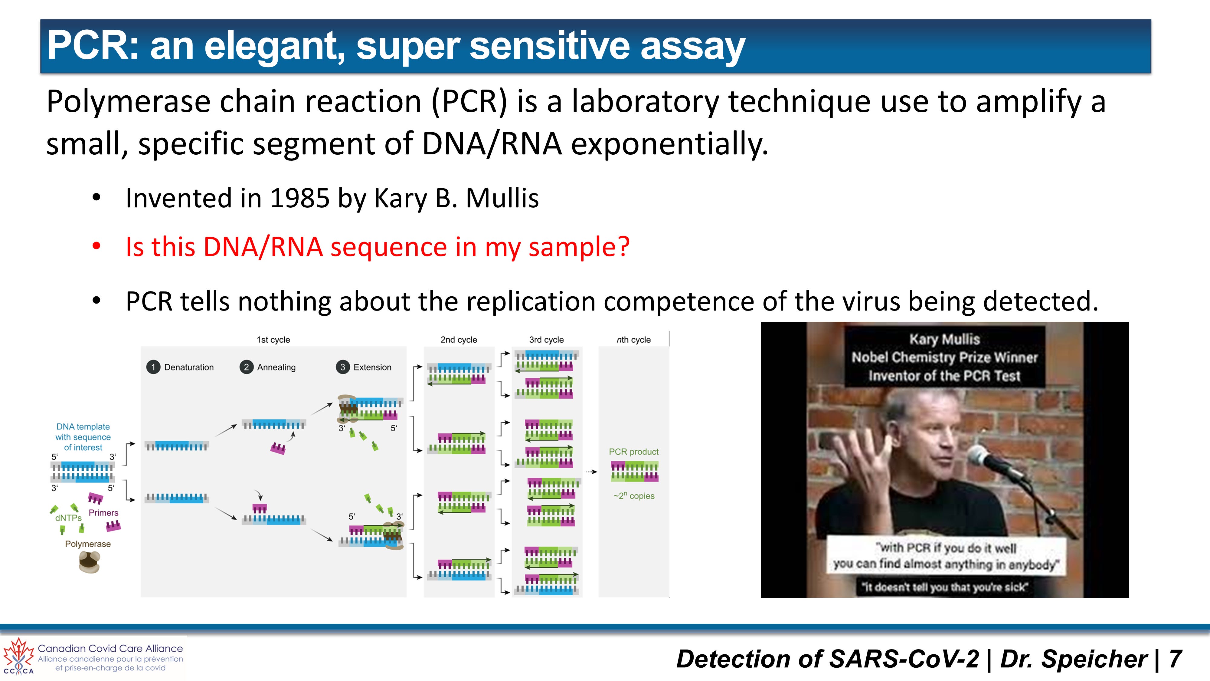
While some people use this quote from Dr Mullis to claim that the assay picks up all DNA sequences in the sample and is not a specific test this is far from reality. I have had some good conversations with Dr Mullis’ bench scientist Steve Scharf. Dr Mullis was a theoretical organic chemist who wasn’t good at doing lab work but was a brilliant mind and worked on the best way to detect and amplify pieces of DNA and RNA. The quote here from Dr Mullis is absolutely correct. If you have properly designed primer sequences you can amplify any piece of known DNA or RNA that you want. This assay only asks “Is this DNA/RNA sequence in my sample?” It does not tell us anything about the replication competence of the virus or if it is the virus that is causing the disease. Therefore, it is critical to have good communication between the attending physician who observes the patient’s sign and symptoms and the clinical molecular laboratory doing the testing.
The Mechanics and Boundaries of Real-Time RT-qPCR
Modern clinical diagnostics utilize real-time Reverse Transcription-quantitative PCR (RT-qPCR). The execution of this assay follows a strict, multi-step pipeline aligned with standard MIQE Guidelines:
Pre-Analytical Collection & Transport: Biological specimens (nasopharyngeal swabs, mid-turbinate swabs, or saliva) are collected into Universal Transport Medium (UTM) and kept under strict temperature controls to prevent the rapid enzymatic degradation of fragile viral RNA.
Extraction & Purification: Automated platforms utilize silica-fiber columns or magnetic beads alongside chaotropic salt buffers and Proteinase K to lyse cellular structures, remove protein inhibitors, and purify total nucleic acids. Human genomic material and viral targets are co-purified during this step.
Reverse Transcription (RT): Since coronaviruses possess an RNA genome, an enzyme called reverse transcriptase converts the fragile viral RNA into complementary DNA (cDNA) at roughly 50°C before amplification can begin.
Thermal Cycling: A thermostable DNA polymerase (derived from Thermus aquaticus, Taq polymerase) executes 30 to 45 automated thermal cycles comprising three steps:
Denaturation (95°C): Unwinding double-stranded cDNA into single strands.
Annealing (50-65°C): Short, highly specific synthetic oligonucleotide primers bind to targeted viral gene sequences (e.g., ORF1ab, N gene, E gene, or RdRP).
Extension (72°C): Taq polymerase synthesizes a new complementary DNA strand.
Fluorescent Detection (TaqMan™ Probes): Dual-labelled hydrolysis probes carry a 5’ reporter fluorophore and a 3’ quencher. As Taq polymerase synthesizes the new strand, its 5’-to-3’ exonuclease activity cleaves the probe, permanently separating the reporter from the quencher and releasing a measurable fluorescent signal.
The cycle at which this accumulating fluorescent signal crosses a fixed background threshold is designated the Cycle Threshold (Ct) or Quantification Cycle (Cq), which one depends on whether or not the assay is used for quantitative analysis. An inverse relationship governs this measurement: a low Ct value indicates a massive starting viral load, whereas a high Ct value means the instrument executed dozens of amplification cycles to detect minute trace amounts of genetic material.
The core scientific boundary of PCR is absolute: PCR detects nucleotide sequences; it does not measure viral replication competence. An RT-qPCR assay functions as a biochemical photocopier. It is biochemically incapable of determining whether the detected target sequence derives from an active, infectious virion or from the non-replicating, degraded genomic “trash” of an infection that the host’s immune system neutralized weeks prior.
The Foundation of Sand: Early Flaws in the CDC Assay Protocol
Before evaluating how PCR was systematically scaled up across healthy populations, we must audit the severe technical, clinical, and regulatory failures that plagued the foundational testing protocols at the start of the pandemic response. As a diagnostic molecular virologist who has managed high-throughput genomic testing facilities, the operational cracks in the system were obvious long before they reached public awareness. The initial rollout of the standard CDC assay protocol—engineered around specific nucleocapsid viral targets—was deeply compromised by structural flaws that established a highly volatile baseline for global diagnostics.
1. The Biochemical Instability of the N2 Target
The standard CDC assay was designed to detect multiple distinct genetic markers within the viral genome, specifically targeting the N1 and N2 regions of the nucleocapsid gene. In the practical environment of high-throughput clinical laboratories, the N2 target proved to be profoundly problematic and biochemically unstable. In early April 2020, laboratory networks on diagnostic channels like ClinMicroNet flagged that the N2 target routinely lagged drastically or failed altogether, crossing the cycle threshold at a Ct value well over the standard 40-cycle cut-off, even when the companion N1 target showed a clean, robust curve. Independent laboratory data, most notably a landmark Yale School of Public Health study (Vogels et al., 2020), confirmed that the CDC’s N2 primer-probe set was uniquely susceptible to background amplification with negative nasopharyngeal swabs, causing systemic false-positive signals and artifacts, whereas the N1 target remained highly stable and specific.
2. The “Inconclusive” Clinical Reporting Trap
The failure of the N2 target triggered an immediate diagnostic and operational crisis. Under the rigid criteria mandated by the initial CDC protocol, a sample could only be reported as a definitive “positive” if both the N1 and N2 targets successfully amplified within designated limits. By April 8, 2020, frontline academic centers—such as the University of Tennessee Health Science Center—warned that clinical samples with strong N1 values but trailing N2 curves were being forced into a dangerous reporting category: “inconclusive.” Detailed internal data evaluations from networks like UCLA Health subsequently revealed that this metric was profoundly misleading. Out of reviewed inconclusive results, over 83% (five out of six) were confirmed via clinical correlation and repeat testing to be true positives capturing patients with lower viral loads—either at the early onset of an active infection or during the late recovery phase. Forcing labs to report these weak positives as “inconclusive” severed laboratory data from clinical reality and delayed proper patient management.
3. Regulatory Whiplash and Institutional Contradiction
As clinical laboratory directors realized that these strict target rules were misclassifying true low-level positive patients, they engaged directly with regulatory bodies to reform the reporting logic. The response from regulators exposed a chaotic internal disconnect. Around April 8, 2020, during an Emergency Use Authorization (EUA) revision process at major institutions like Memorial Sloan Kettering Cancer Center, the FDA Hub explicitly instructed clinical laboratories to alter their interpretation criteria, advising them to report samples as definitively “positive” when either the N1 or the N2 target alone was detected. Yet, in an act of staggering institutional whiplash on April 9, 2020, the FDA abruptly rescinded this recommendation. The regulatory agency backtracked, telling labs to completely ignore their previous directive simply because the CDC had not yet officially altered its own baseline assay interpretation document. Clinical labs were left stranded between clinical common sense and rigid regulatory compliance.
4. Administrative Barriers to Real-Time Scientific Optimization
This regulatory gridlock created an artificial barrier that penalized scientific accuracy and suppressed frontline laboratory expertise. When laboratory directors attempted to implement common-sense clinical modifications in April 2020—such as interpreting an inconclusive result as a weak positive if the patient presented with overt symptoms (e.g., fever, cough, shortness of breath, chest imaging findings) or a known exposure history—they were met with administrative threats. The FDA signaled that any permanent change to the interpretation criteria of the validated CDC assay would require a brand-new, entirely independent EUA submission. By transforming a routine clinical interpretation update into an arduous, prohibitive administrative hurdle, regulatory frameworks actively choked off real-time optimization, locking clinical laboratories into a flawed diagnostic framework that systematically distorted the true epidemiological picture.
Dr Speicher’s May 2023 NCI Testimony: Deconstructing the Eight Misinformation Points & The “Too Many Cycles” Myth
During my time directing clinical diagnostic laboratories, it became glaringly obvious that public health agencies were conflating analytical detection with clinical disease. During my May 2023 NCI expert witness testimony, I systematically dismantled eight pervasive points of misinformation surrounding PCR testing.

Watch my NCI Testimony here: https://nationalcitizensinquiry.ca/witness/dr-david-speicher/
Misinformation #1: Speech Fluency Defines Intellect and Authority
Opening my very first public presentation of the pandemic with this point was both intentional and deeply personal. Public perception often falsely associates speech impediments with reduced intellectual capacity or personal insecurity. Navigating a lifelong stutter has proven to be a profound strength throughout my career. Standing before parliamentary committees, courtrooms, and national inquiries with a deliberate, methodical, and unhurried cadence reflects deep conviction. Pacing reflects purpose. True authority is defined by empirical truth and moral fortitude, not polished rhetoric designed to deceive.
Misinformation #2: PCR Results are 97% False Positives and Should Be Abandoned Entirely
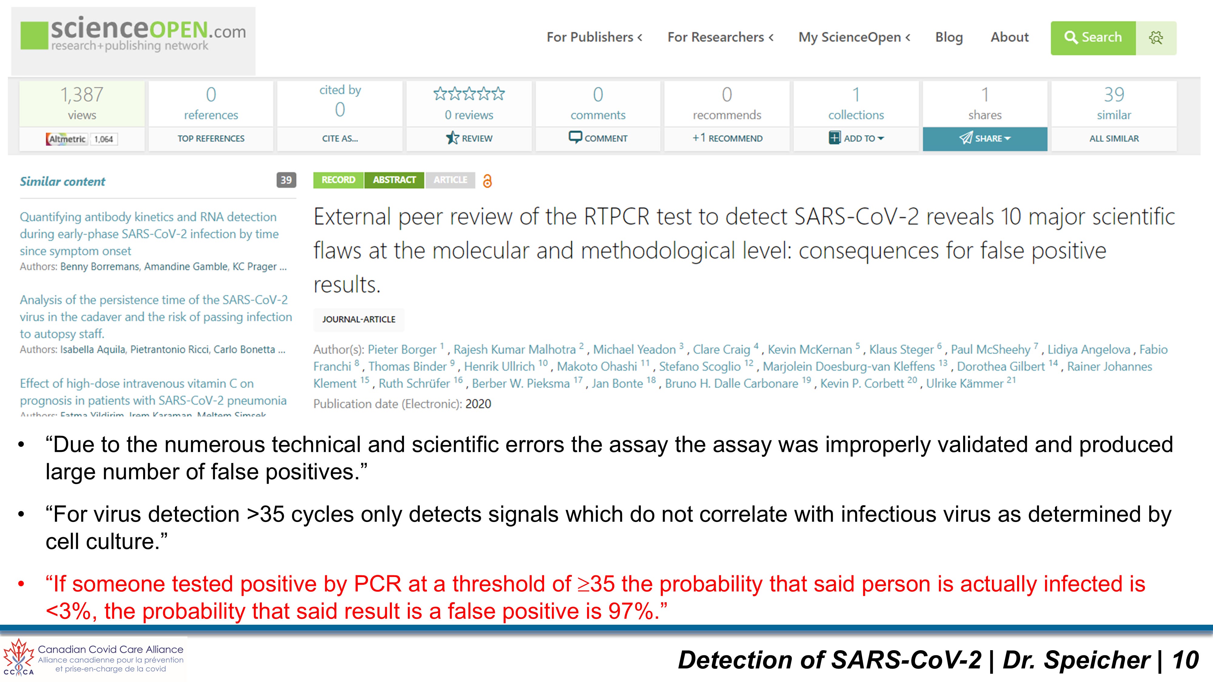
Pervasive claims asserting that PCR testing is inherently fraudulent or “97% false positive” misrepresent foundational diagnostic science. This figure, derived from landmark reviews such as Borger et al. (2020), establishes that when an individual tests positive by PCR at a high threshold Ct>35, the probability that the individual harbours active, replication-competent virus is under 3%. The remaining 97% represents a false positive for clinical infectivity.
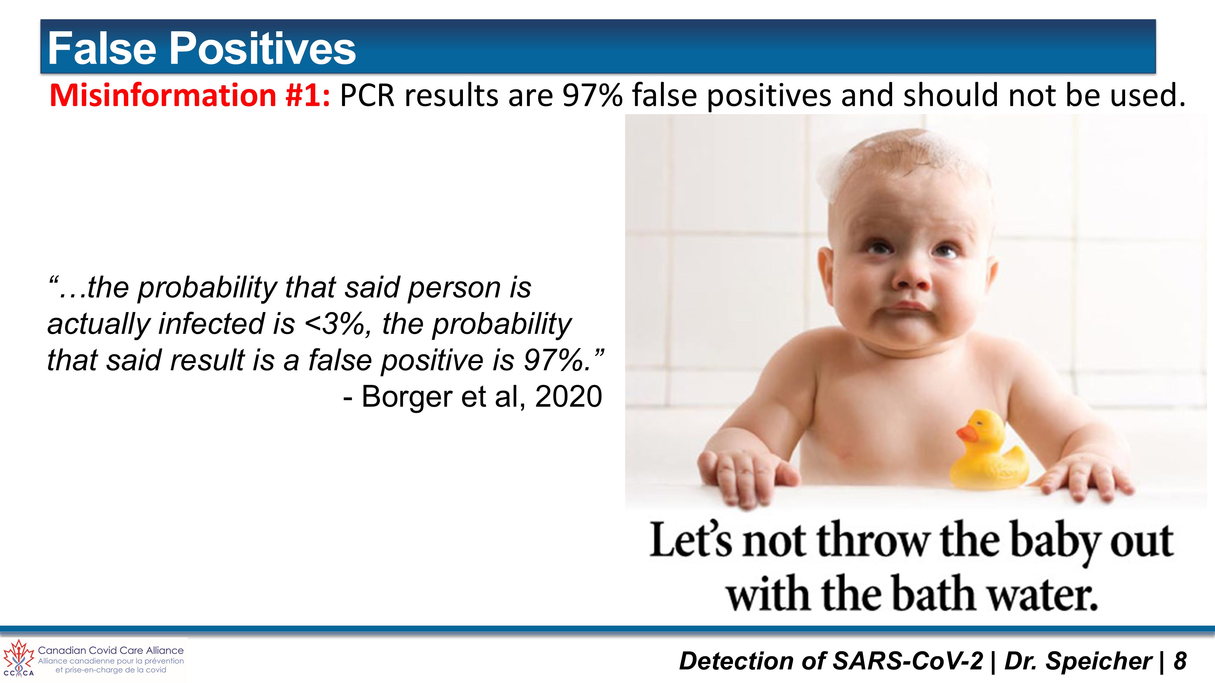
This statistic does not mean the PCR instrument suffered a biochemical failure or generated an imaginary signal. The fluorescent probe bound accurately to the target nucleotide sequence, confirming analytical presence. The error lies in substituting analytical detection for clinical diagnosis. In other words, the difference betweent he assays analytical limit of detection and the clinical limit of detection (i.e. the lowest viral load required to cause clinical illness). At Ct=35, the target sequence almost universally represents non-infectious viral debris—shattered genomic remnants leftover from a cleared infection—or background laboratory contamination.
Dismissing PCR entirely due to this misapplication constitutes “throwing the baby out with the bathwater”. Such dismissal undermines the very technology used to expose residual plasmid DNA and SV40 promoter-enhancer sequences in modRNA vaccines. The failure was not in the chemistry of the assay, but in the reckless public health decision to treat late-cycle analytical signals in healthy individuals as active clinical “cases”.
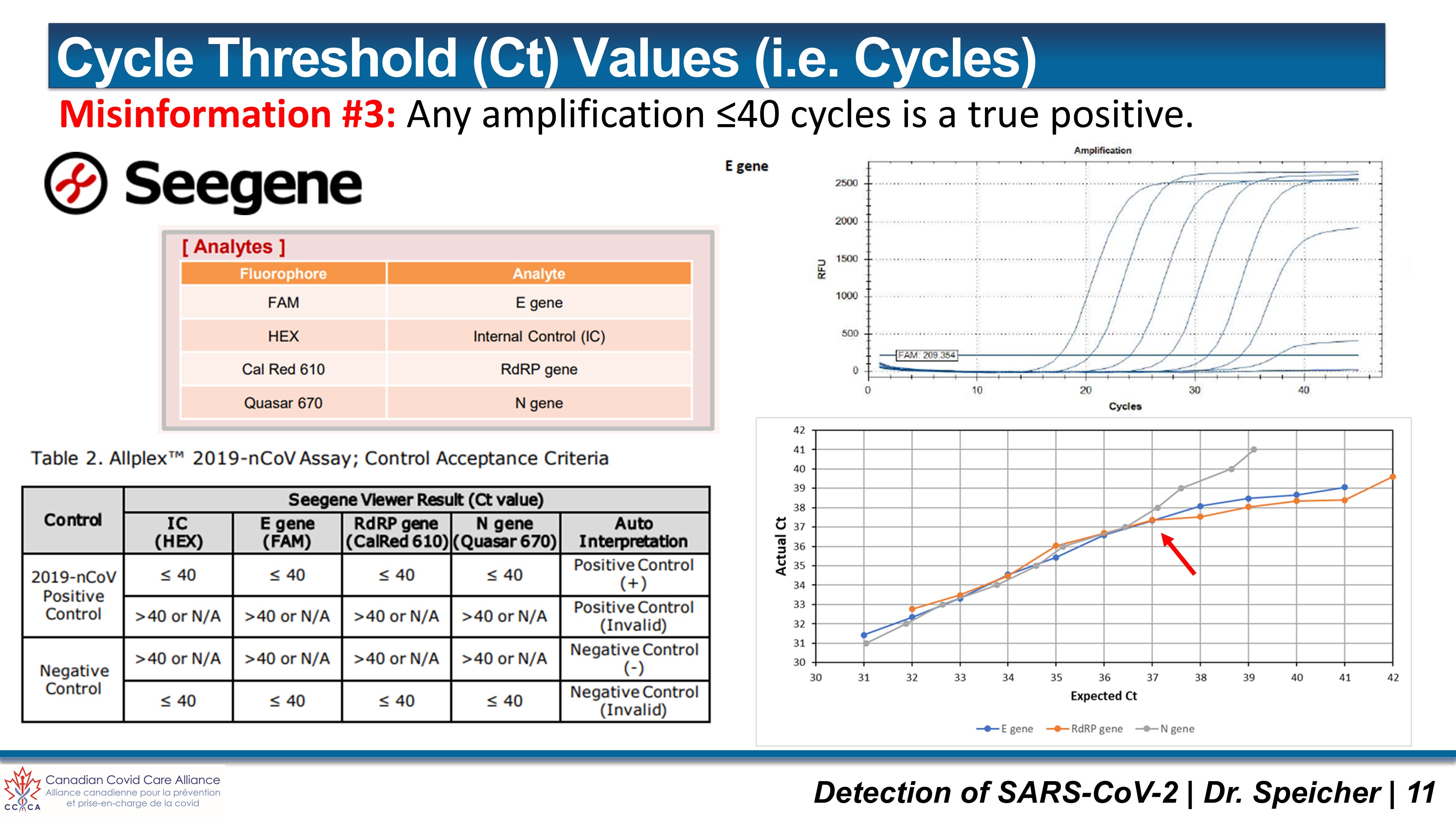
Misinformation #3: The “Too Many Cycles” Myth vs. Clinical Cut-Off Reality
A widespread public and scientific misconception asserts that simply running a PCR machine for 40 to 45 cycles is inherently erroneous. This reflects a fundamental misunderstanding of laboratory science. Running a real-time PCR assay out to 40 or 45 cycles is standard scientific practice; it is necessary to establish a proper fluorescence baseline and accurately observe what occurs at the assay’s Limit of Detection (LOD).
The diagnostic error lies not in how many cycles the machine is programmed to run, but where the clinical cut-off between a “positive” and a “negative” result is set.
A highly specific fluorescent probe (such as TaqMan™) will bind to a shattered, non-infectious fragment of viral RNA at cycle 38 and emit a signal. That signal merely proves the machine accurately detected the biological artifact it was engineered to find. Viral culture studies (e.g., Bullard et al., 2020) prove that replication-competent, infectious virus cannot be isolated from specimens with Ct values exceeding 24 to 27. Beyond 30 cycles, viral load is simply too low to consistently cause a cytopathic effect in cell culture, making live virus isolation practically impossible.
A single static high Ct value represents a profound diagnostic ambiguity. As noted by expert clinical microbiologists such as Dr. Vickie Baselski, a low-level fluorescent signal does not exclusively signify a historical artifact or non-replicating genomic trash from a cleared infection. It can equally represent an emerging viral load in the early pre-symptomatic incubation phase of an active infection. During this early incubation period, the host is in a state of rapid, deterministic exponential viral replication. Far from being harmless genomic debris, an individual caught at the very onset of viral synthesis is on the verge of becoming highly infectious and poses an active biological risk.
This dual-directional reality demonstrates precisely why a standalone, isolated Ct value cannot serve as an algorithmic proxy for public health policy. Without clinical correlation—evaluating symptom onset, exposure history, and conducting serial re-testing to observe whether the cycle threshold is rapidly decreasing (emerging infection) or remaining stably elevated (cleared remnant)—a static laboratory result is incapable of distinguishing between an ascending threat and a harmless biological echo.
Clinical microbiology requires two distinct, non-negotiable cut-offs:
Assay Limit of Detection (LOD): Ct 35–37 (detecting trace nucleic acid fragments or early incubation).
Clinical Infectious Dose Threshold: Ct 30 (indicating a viral load capable of active transmission and clinical illness).
Reporting late-cycle signals Ct > 35 as active clinical cases without serial testing or physical examination ignored the assay’s validated LOD and transformed degraded genomic trash into public health panics.
Misinformation #4: Test Results and Ct Values Can Be Direct Comparison Metrics Between Different Laboratories
Cycle threshold values are not absolute, standardized units of measure. A Ct count generated on one laboratory platform cannot be directly compared to a Ct count from another facility. Raw cycle values fluctuate drastically based on a wide array of pre-analytical and analytical variables:
Clinical & Testing History: Patient symptom duration, timing relative to exposure, and previous infection status.
Sample Matrix & Collection Depth: Nasopharyngeal swab vs. mid-turbinate nasal swab vs. oral swab vs. self-collected saliva.
Transport Media & Cold Chain: Volume of Universal Transport Medium (UTM) and temperature management during transport.
Extraction Efficiency & Chemistry: Silica-column vs. magnetic bead automated extraction kinetics.
Instrumentation & Calibration: Thermocyler model, optical channel calibration, and fluorescence threshold setting.
Assay Design & Targets: Specific gene targets (ORF1ab, N, E, RdRP) and primer/probe binding efficiency.
Daily Run Performance: Reagent batch variation and daily background control levels.
Every accredited laboratory is required to independently validate its specific assays and establish its own internal cut-off limits based on its unique instrumentation and workflows.
Crucially, in routine healthcare delivery, administering physicians are not provided with the raw Ct value. Diagnostic laboratory reports delivered to clinicians state only a binary call: “Detected / Positive” or “Not Detected / Non-Reactive” (or occasionally “Inconclusive”).
Withholding raw Ct values is a deliberate structural practice in laboratory medicine. Most practicing physicians have not been trained in molecular diagnostic kinetics to interpret raw cycle counts in isolation without laboratory context, and without knowing the specific testing platform’s Limit of Detection (LOD). Interpreting a raw Ct value requires deep technical knowledge of the specific instrument, extraction protocol, and assay baseline.
This communication gap creates a severe clinical disconnect. A physician reviewing a patient report sees the exact same “Positive” label whether the patient has a massive, highly infectious viral load (Ct = 18) or carries low levels of non-infectious genomic debris from a cleared infection three months prior (Ct = 36).
This reality underscores the vital need for stronger, collaborative partnerships between administering physicians and clinical diagnostic microbiologists. Laboratory data should never be used as an automated, standalone directive. Diagnostic results must be interpreted through direct dialogue between the clinician evaluating the patient’s physical presentation and the diagnostic laboratory director who understands the analytical kinetics of the assay.
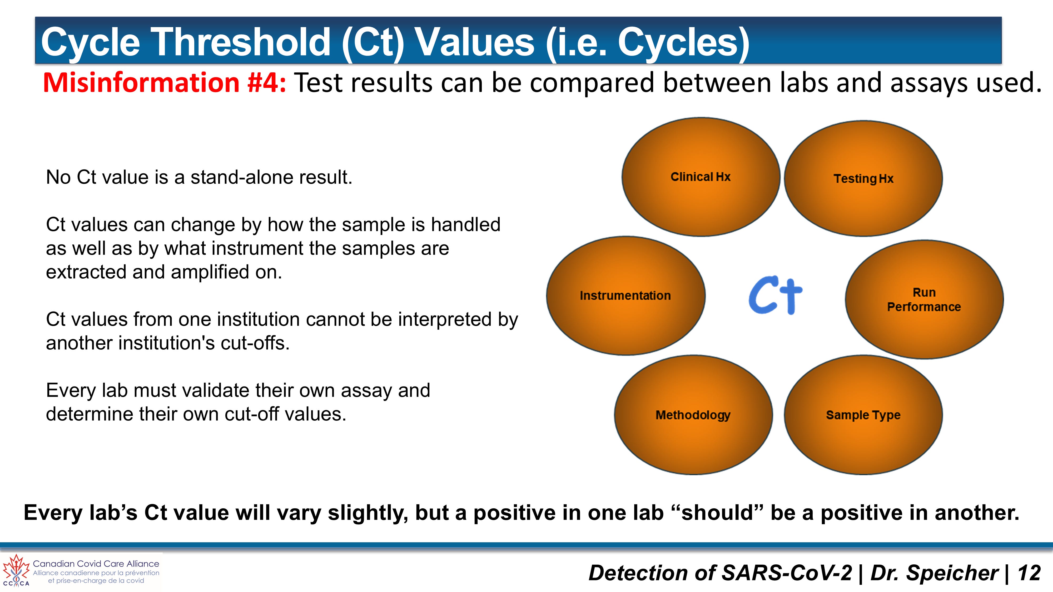
Misinformation #5: Invasive Nasopharyngeal Swabbing is Mandatory for Accurate Viral Detection
Public health directives insisted that deep, invasive nasopharyngeal (NP) swabbing was the sole acceptable collection matrix for accurate viral detection. NP collection requires inserting a flexible mini-tip swab deep into the nasal cavity—parallel to the palate at a 70° head tilt until resistance is met at the posterior pharynx—to scrape epithelial cells. While NP swabs remain the clinical gold standard for hospitalized patients requiring high analytical sensitivity (97.85%), mandating this invasive procedure across millions of healthy citizens created severe operational bottlenecks and unnecessary discomfort.
A comprehensive evaluation of alternative diagnostic matrices demonstrates that less invasive sampling methods deliver exceptional diagnostic performance for detecting active viral shedding:
Nasopharyngeal (NP) Swabs: Highest diagnostic sensitivity (97.85%; 95% CI: 94.62–100%). Optimal for low viral loads (Ct > 30), but highly invasive, requiring trained healthcare personnel and specialized personal protective equipment (PPE).
Mid-Turbinate (MT) & Anterior Nasal Swabs (NS): Minimally invasive, swabbing the inferior turbinate or nostrils. Overall sensitivity sits at roughly 87.10% (95% CI: 79.57–93.55%). Crucially, diagnostic concordance with NP swabs reaches 94% when viral loads are high (Ct <30). Concordance drops to 41% only at low viral loads (Ct > 30), proving that MT swabs reliably capture all clinically infectious individuals while filtering out non-infectious background signals.
Oral Flocked Swabs & Combined Throat/Nares Swabs: Highly effective non-invasive alternatives. As documented in studies I co-authored (Speicher et al., 2020), oral flocked swabs provide robust yield for viral nucleic acids and mucosal antibodies while significantly reducing swab discomfort and specimen collection friction.
Self-Collected Saliva (Neat or Swish-and-Gargle): Entirely non-invasive, cost-effective, and collectable without direct clinical intervention. In our landmark systematic review (Khurshid, Zohaib... Speicher DJ, 2020), we established that saliva achieves an overall diagnostic sensitivity of 87.10% (95% CI: 79.57–93.55%). When paired with a simple nasal swab (NS + Saliva), combined sensitivity reaches 94.62% (95% CI: 89.25–98.92%).
For population-level screening or identifying individuals actively shedding replication-competent virus, non-invasive matrices—specifically saliva and oral/nasal swabs—provide diagnostic equivalence to NP swabbing without subjecting citizens to unnecessary physical coercion.
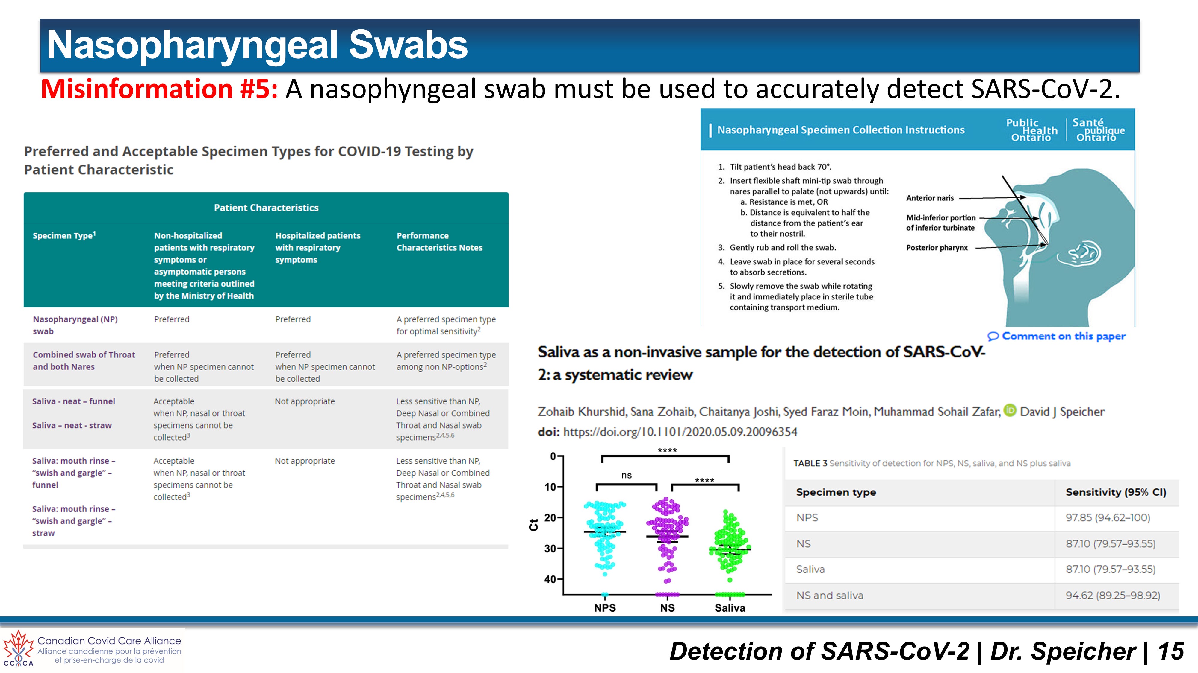
Misinformation #6: Every PCR-Positive Individual Represents a Sick “Case” of COVID-19
Classical clinical medicine requires three distinct terms to be accurately defined:
Infected: An individual harboring viral nucleic acid or non-replicating fragments, often entirely asymptomatic and non-infectious.
Infectious: An individual harboring a sufficient active, replicating viral load to shed live virus and infect others.
Diseased: An individual experiencing overt clinical symptoms and biological illness.
In February and March 2020, during the early development of in-house PCR assays at St. Joseph’s Healthcare Hamilton, I observed this foundational disconnect firsthand. I asked a Medical Microbiology and Infectious Disease physician why the laboratory was labeling positive PCR test results as “COVID” positive rather than “SARS-CoV-2” positive. The doctor replied that doing so was simply “easier and less confusing for physicians.”
In hindsight, this semantic substitution carried catastrophic public health consequences. SARS-CoV-2 is the target virus; COVID-19 is the clinical disease. Labelling every positive viral RNA detection as a “COVID case”—regardless of whether the patient presented with a fever, cough, or zero symptoms whatsoever—severed diagnostic testing from clinical evaluation. Mass testing protocols blurred these boundaries completely. Millions of healthy, asymptomatic individuals shedding microscopic, non-replicating viral debris were classified as active “cases” of COVID-19. Reporting case counts based exclusively on positive PCR signals without physical presentation manufactured an artificially inflated metric of threat that drove public panic and dictated destructive policy decisions.
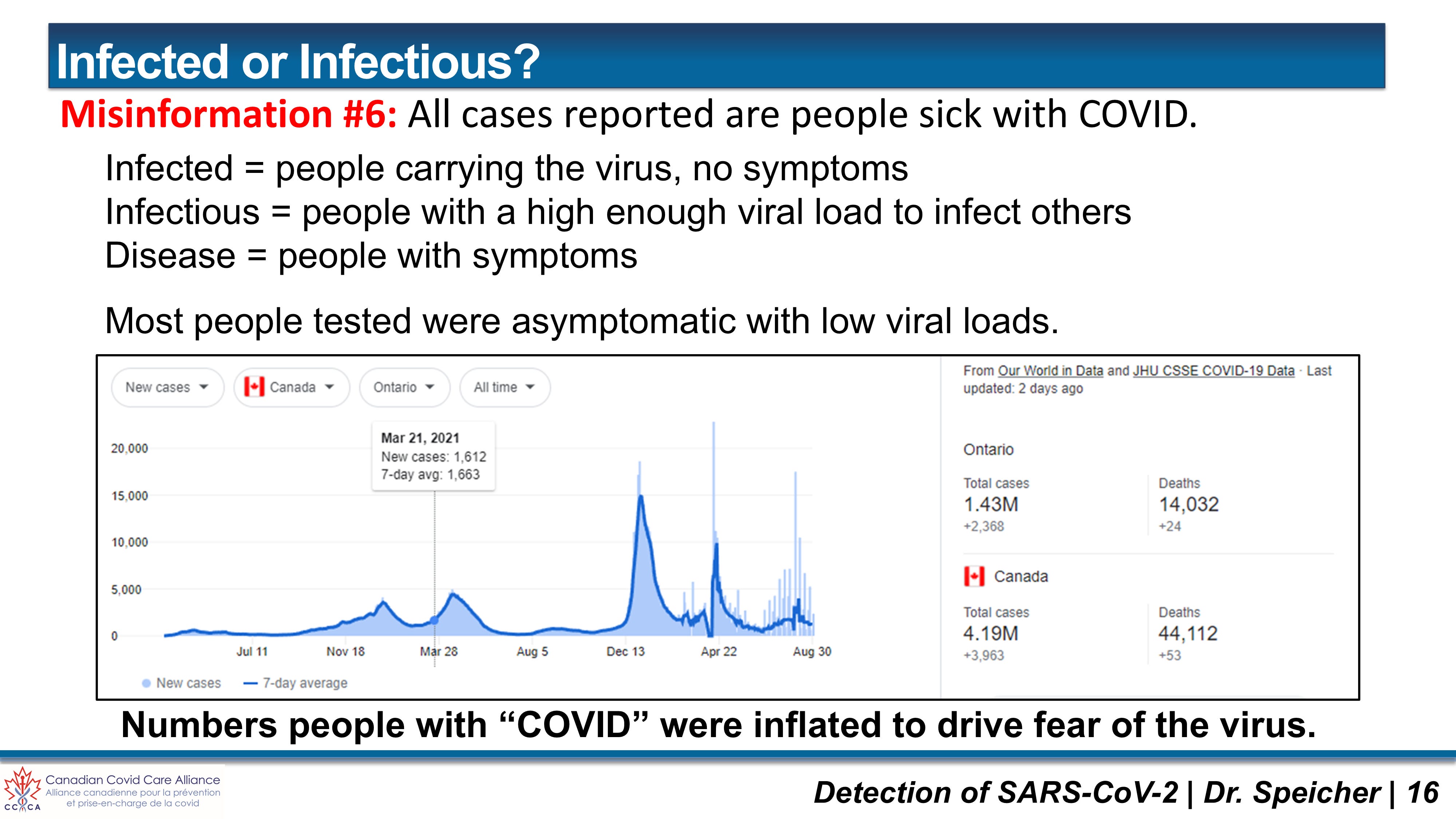
Misinformation #7: Asymptomatic Transmission is a Primary Driver of Pandemic Spread
The narrative asserting that healthy, symptom-free individuals were silently driving pandemic transmission defied foundational epidemiology. Achieving viral transmission requires an Infectious Dose (ID50) of roughly 500 to 2,000 replication-competent virions—a viral load that almost universally produces active clinical symptoms. Large-scale epidemiological studies confirmed that true asymptomatic transmission is exceptionally rare. Individuals recovering from an infection can continue shedding dead, non-infectious viral fragments detectable by high-cycle PCR for up to 90 days. Non-infectious debris cannot drive an outbreak.
Misinformation #8: Rapid Antigen Tests (RATs) are Cheap, Quick Screening Tools for Asymptomatic Populations
Rapid Antigen Tests (RATs)—such as the Abbott Panbio™ and BNTX Rapid Response™—are lateral flow immunoassays engineered to detect specific viral structural proteins, primarily the nucleocapsid (N) protein, via direct antibody binding on a nitrocellulose strip. Unlike PCR, RATs possess zero biochemical amplification mechanisms; their visual readout depends entirely on the physical concentration of target antigen present in the primary biological specimen.
Public health authorities and corporate entities deployed RATs en masse as rapid, cheap gatekeeping mechanisms to screen millions of healthy, asymptomatic cohorts. This mass deployment represented a fundamental misuse of diagnostic technology. A lateral flow immunoassay without amplification is mechanically incapable of reliably detecting low viral loads. While RATs perform acceptably during peak symptomatic infection when viral loads are massive (Ct <25), systematic evaluations—such as those published by the Ontario COVID-19 Science Advisory Table (Jüni et al., 2021)—confirm that RAT sensitivity degrades precipitously to 20.9% or lower in specimens with Ct >30. Deploying RATs across healthy cohorts with low pre-test probability generated widespread false reassurance through false negatives, while simultaneously triggering false-positive panics due to the biochemical volatility of the devices—where minor fluctuations in specimen pH (below 7.0) or buffer Tricine concentration cause non-specific binding.
Health Canada and global regulatory agencies explicitly classified RAT results as presumptive only, mandating that any positive test be confirmed via quantitative PCR. Bypassing this clinical mandate and utilizing RATs as standalone determinants of employment or social participation was scientifically indefensible.
The financial and logistical cost of this policy failure was staggering. Governments procured hundreds of millions of rapid test kits at public expense, allocating hundreds of millions of taxpayer dollars to private distributors and diagnostic middlemen. Millions of these test kits expired in warehouses, were distributed indiscriminately to uninterested workplaces, or were discarded unused into landfills—literally flushing public capital down the drain. Taxpayer funds that should have fortified frontline clinical care, supported vulnerable citizens, and funded transparent, independent safety auditing were instead squandered on fragile plastic cassettes that provided an illusory sense of biosecurity.
The Imperative of Clinical Integration & Modern Diagnostic Frontiers
Modern healthcare requires robust, highly sensitive, and cost-effective diagnostic tools. An excellent diagnostic test must deliver rapid turnaround times, exquisite specificity, and actionable clinical clarity without imposing prohibitive financial burdens on healthcare infrastructure. The ultimate purpose of a diagnostic test, however, is not to replace clinical judgment, but to serve as a supportive instrument alongside physical bedside evaluation.
Diagnosing a patient solely on an isolated laboratory readout—severed from physical clinical evaluation—violates fundamental medical principles. A positive laboratory signal is a biological data point, not an automated clinical diagnosis. An administering physician evaluating a patient at the bedside observes physical signs: fever, respiratory distress, auscultatory crackles, or systemic inflammation. The clinical diagnostic laboratory must work in direct, collaborative alignment with the physician, providing the molecular or serological context required to refine the differential diagnosis and confirm the underlying pathogen. Substituting physical examination with automated nucleic acid amplification manufactured a “casedemic”—an artificially inflated metric of disease driven by positive laboratory signals rather than actual sick patients.
As we advance toward new diagnostic horizons, molecular medicine is actively evolving to resolve the historical constraints of conventional assays. Two major technological advancements illustrate this paradigm shift:
MultiSeq™ Technologies (Allele-Specific Multiplex Sequencing): Developed to bridge the gap between high-sensitivity qPCR screening and definitive Next-Generation Sequencing (NGS), MultiSeq™ utilizes Allele-Specific Multiplex Sequencing (ASMS) to integrate target amplification and sequence confirmation into a single, six-hour workflow. By modifying chain-termination chemistry, MultiSeq™ simultaneously detects multiple overlapping pathogens, confirms specific species, and identifies critical antimicrobial resistance (AMR) markers (such as mecA in MRSA) in a single test well. This eliminates the financial and temporal burden of secondary “reflex” testing, providing sequence-verified diagnostic certainty directly to the treating physician.
LIDAR qPCR (Longitudinal ID of Amplified Reporters): Pioneered by Kevin McKernan and Medicinal Genomics (McKernan, 2026), LIDAR qPCR resolves the long-standing trade-off between SYBR Green intercalating dyes and TaqMan™ hydrolysis probes. Standard TaqMan™ assays require a long target sequence footprint (>75bp) and undergo irreversible 5’-to-3’ exonuclease cleavage during amplification, preventing subsequent High-Resolution Melt (HRM) analysis and under-reporting fragmented DNA by 16- to 32-fold. LIDAR qPCR utilizes universal 5’ Fluorophore-Quencher “Probers” paired with unlabeled bridge primers. When the expanding DNA helix forces the strand into a double-stranded conformation, the fluorophore and quencher separate by 3.4 Å per base, producing a reversible 5-color fluorescent signal. Because LIDAR consumes zero internal template footprint, it enables 5-color multiplexed HRM analysis on ultra-short DNA targets (50-60bp) without probe destruction. This breakthrough enables high-precision quantification and sequence-context profiling of highly fragmented DNA—such as residual plasmid contaminants in modRNA vaccines, circulating tumor DNA (ctDNA), and ancient DNA—delivering a direct, 1st-step readout of PCR kinetics at a fraction of standard probe synthesis costs. Read Kevin McKernan’s full LIDAR analysis here.
We are called to be salt and light in an environment that frequently prefers the safety of silence. My technical testimony before the National Citizens Inquiry was delivered to ensure that the scientific facts regarding molecular testing were permanently etched into our national legal record. As we prepare to address new technological frontiers—including the regulatory oversight of modRNA platforms, residual DNA contamination, and synthetic biology at the upcoming August 2026 NCI hearings—our mandate remains unchanged.
We must stand firm on a foundation of scientific integrity, demanding absolute transparency, rigorous laboratory standards, and the protection of human dignity. Stand with us in this ongoing effort. Share these data-driven analyses, support independent scientific auditing, and subscribe to join our pursuit of unvarnished truth.
References
Borger, P., Malhotra, R. K., Yeadon, M., Craig, C., McKernan, K., Steger, K., McSheehy, P., Angelova, L., Franchi, F., Binder, T., Ullrich, H., Ohashi, M., Scoglio, S., Doesburg-van Kleffens, M., Gilbert, D., Klement, R. J., Schrüfer, R., Pieksma, B. W., Bonte, J., Dalle Carbonare, B. H., Corbett, K. P., & Kämmerer, U. (2020). External peer review of the RTPCR test to detect SARS-CoV-2 reveals 10 major scientific flaws at the molecular and methodological level: consequences for false positive results. ScienceOpen. https://doi.org/10.32945/osf.io/9yx7q
Bullard, J., Dust, K., Funk, D., Strong, J. E., Alexander, D., Garnett, L., Beda, C., Ciurysek, L., Poliquin, G., Zarychanski, R., Rusk, N., Tran, K. N., Liang, B., Wood, H., Mahmood, R., Nelson, A., & Head, M. M. (2020). Predicting infectious Severe Acute Respiratory Syndrome Coronavirus 2 from diagnostic samples. Clinical Infectious Diseases, 71(10), 2663–2666. https://pubmed.ncbi.nlm.nih.gov/32442256/
Bustin, S. A., Benes, V., Garson, J. A., Hellemans, J., Huggett, J., Kubista, M., Mueller, R., Nolan, T., Pfaffl, M. W., Shipley, G. L., Vandesompele, J., & Wittwer, C. T. (2009). The MIQE guidelines: minimum information for publication of quantitative real-time PCR experiments. Clinical Chemistry, 55(4), 611–622. https://doi.org/10.1373/clinchem.2008.112797
Corman, V. M., Landt, O., Kaiser, M., Molenkamp, R., Meijer, A., Chu, D. K., Bleicker, T., Brünink, S., Schneider, J., Schmidt, M. L., Mulders, D. G., Haagmans, B. L., van der Veer, B., van den Brink, S., Wijsman, L., Goderski, G., Romette, J. L., Ellis, J., Zambon, M., Peiris, M., Goossens, H., Reusken, C., Koopmans, M. P., & Drosten, C. (2020). Detection of 2019 novel coronavirus (2019-nCoV) by real-time RT-PCR. Eurosurveillance, 25(3), 2000045. https://doi.org/10.2807/1560-7917.ES.2020.25.3.2000045
Jüni, P., Baert, S., Bobos, P., et al. (2021). Rapid antigen tests for voluntary screen testing. Science Briefs of the Ontario COVID-19 Science Advisory Table, 2(52). https://doi.org/10.47326/ocsat.2021.02.52.1.0
Khurshid, Z., Zohaib, S., Joshi, C., Moin, S. F., Zafar, M. S., & Speicher, D. J. (2020). Saliva as a non-invasive sample for the detection of SARS-CoV-2: a systematic review. medRxiv. https://doi.org/10.1101/2020.05.09.20096354
McKernan, K. (2026, July 18). A novel qPCR detection chemistry to address the quantitation of fragmented DNA populations: LIDAR. Nepetalactone Newsletter (Substack).
Mullis, K., Faloona, F., Scharf, S., Saiki, R., Horn, G., & Erlich, H. (1986). Specific enzymatic amplification of DNA in vitro: the polymerase chain reaction. Cold Spring Harbor Symposia on Quantitative Biology, 51(Pt 1), 263–273. https://doi.org/10.1101/sqb.1986.051.01.032
National Citizens Inquiry (NCI). (2023). National Citizens Inquiry Commissioners’ Report. Authored by K. Drysdale, H. DiGregorio, J. Kaikkonen, & B. Massie. https://nationalcitizensinquiry.ca/commissioners-report/
Pinninti, S., Trieu, C., Pati, S. K., et al. (2020). Comparing nasopharyngeal and mid-turbinate nasal swab testing for the identification of SARS-CoV-2. Clinical Infectious Diseases, 72(11), 1981–1983. https://doi.org/10.1093/cid/ciaa882
Public Health Ontario (PHO). (2020). FOCUS ON: An overview of cycle threshold values and their role in SARS-CoV-2 real-time PCR test interpretation. Toronto, ON: Queen’s Printer for Ontario.
Speicher, D. J. (2023, May 18). Detection of SARS-CoV-2 by PCR: Misuse of an Elegant Technique. Expert Witness Testimony presented at the National Citizens Inquiry (NCI), Ottawa, ON.
Speicher, D. J., Ali, M., & Smieja, M. (2016). Respiratory viruses. In Manual of Molecular and Clinical Laboratory Immunology (8th ed., Chapter 63, pp. 598–609). ASM Press. https://doi.org/10.1128/9781555818722.ch63
Speicher, D. J., Luinstra, K., Smith, E. J., Castriciano, S., & Smieja, M. (2020). Non-invasive detection of IgG antibodies from common pathogenic viruses using oral flocked swabs. Diagnostic Microbiology and Infectious Disease, 97(3), 115038. https://doi.org/10.1016/j.diagmicrobio.2020.115038
Speicher, D. J., Rose, J., & McKernan, K. (2025). Quantification of residual plasmid DNA and SV40 promoter-enhancer sequences in Pfizer/BioNTech and Moderna modRNA COVID-19 vaccines from Ontario, Canada. Autoimmunity, 58(1), 2551517. https://doi.org/10.1080/08916934.2025.2551517
Vogels, C. B. F., Brito, A. F., Wyllie, A. L., Fauver, J. R., Ott, I. M., Kalinich, C. C., Petrone, M. E., Casanovas-Massana, A., Muenker, M. C., Moore, A. J., Klein, J., Lu, P., Lu-Culligan, A., Jiang, X., Djurisic, M., White, E. B., Lapidus, S., Kalodimos, H. C., Grubaugh, N. D., & Ko, A. I. (2020). Analytical sensitivity and efficiency comparisons of SARS-CoV-2 RT-qPCR primer-probe sets. Nature Microbiology, 5(10), 1299–1305. https://doi.org/10.1038/s41564-020-0761-6
Support Independent Scientific Oversight
Life-saving diagnostic data, molecular kinetics, and rigorous regulatory due diligence must never be hidden behind an institutional firewall. All of our core PCR validation frameworks, peer-reviewed data, and diagnostic risk assessments will always remain free and accessible to the public.
Conducting uncompromising molecular auditing and dismantling narrative-driven “casedemics” requires a heavily resourced war chest. True independent science demands independent funding. Capitalize our ongoing laboratory operations, physical facility expansion, and public health advocacy by upgrading to a paid Substack subscription today. Your backing ensures we can build health policies on a firm foundation of empirical truth—keeping this technical intelligence weaponized for the clinicians, litigators, and independent lawmakers who need it most.
Deploy the Blueprint & Hold the Line
Share the Evidence: The architects of the prevailing public health narrative rely on public isolation and diagnostic illiteracy. Do not let them control the conversation. Distribute this forensic analysis, the RT-qPCR kinetics breakdown, and our peer-reviewed literature to your professional networks, clinical colleagues, and elected representatives.
Join the Conversation: How do we restore physical clinical evaluation to molecular microbiology and permanently prevent diagnostic instruments from being weaponized to manufacture “casedemics”? What regulatory safeguards are required to enforce a strict boundary between analytical Limit of Detection (Ct >35) and clinical infectivity (Ct <30)? Log your technical and legal assessments in the comments below. We are building a permanent record.
Strategic Funding: To directly resource our independent genomic laboratory space outside of captured institutional platforms, direct contributions can be made via e-transfer support@davidspeicher.com or through my active GiveSendGo campaign.
B2B Advisory & Litigation Defense (Cyrus Scientific Inc.): I operate exclusively on a retained executive basis. For Am Law 100 mass-tort litigators, federal regulatory reformists, and clinical testing networks requiring uncompromising technical due diligence, molecular assay validation, or high-stakes expert-witness defense, direct your formal inquiries to research@davidspeicher.com.
Monitor the Intelligence: Follow my real-time updates on X (@DJSpeicher) and track Cyrus Scientific and Dr. David Speicher on LinkedIn.
Science at it's best! Thank you David for your ongoing hard work!!!
That was a lot of good info. Will need t read that a few times.